Neck Muscle Diagram Labeled - Superficial Veins And Cutaneous Nerves Of Neck - The human body has three different types of muscles.. Why is it important to learn muscle anatomy? The muscles of the human body can be categorized into a number of groups which include muscles relating to the head and neck, muscles of the torso or trunk, muscles of the upper limbs, and muscles of the lower limbs. Muscle and anatomy are two words that are often heard when you are studying science. Contain the common carotid artery, internal. Muscles of the neck (musculi cervicales) the muscles of the neck are muscles that cover the area of the neck hese muscles are mainly responsible for the movement of the head in all directions they consist of 3 main groups of muscles:
The content of the neck is grouped into 4 neck spaces, called the compartments. For more anatomy content please follow us and visit our. Contains glands ( thyroid, parathyroid, and thymus ), the larynx, pharynx and trachea. Anterior, lateral and posterior groups, based on their position in the neck.the musculature of the neck is further divided into more specific groups. Related posts of anatomy of neck muscles diagram abdominal anatomy musclse.
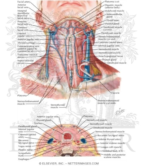
Anatomy Of The Thyroid And Parathyroid Glands Superficial Veins And Cutaneous Nerves Of Neck from netterimages.com Many in the neck help to stabilize or move the head. The neck muscles including the sternocleidomastoid and the trapezius are responsible for the gross motor movement in the muscular system of the head and neck. There are several important vascular structures within the anterior triangle. Broadly considered, human muscle—like the muscles of all vertebrates—is often divided into striated muscle, smooth muscle, and cardiac muscle. The neck is one of the most complex and intricate structures in our body and includes the spinal cord, which sends messages from the brain to the rest of the body. Here is a list of the many muscles that exist in the neck. Labeled pectoralis transversus, and the pectoantebrachialis is labeled pectoralis descendens. Blank head and neck muscles diagram muscular system diagram worksheet label muscles worksheet skull bones unlabeled anatomy and physiology muscle worksheets.
The muscles of the neck run from the base of the skull to the upper back and work together to bend the head and.
Use the memorization method that works best for you. Contains glands ( thyroid, parathyroid, and thymus ), the larynx, pharynx and trachea. Larynx, neck, and lower neck muscles; Causes of neck pain and how to manage the pain in basic terms, the neck (cervical spine) joins the shoulders and chest to the head. 10 best printable worksheets muscle anatomy. Muscle and anatomy are two words that are often heard when you are studying science. The thyroid gland is one of the largest endocrine glands in the body. Broadly considered, human muscle—like the muscles of all vertebrates—is often divided into striated muscle, smooth muscle, and cardiac muscle. Just need a glimpse, leave your valuable advice let us know , and subscribe us! The muscles in this part of the neck are divided as to where they lie in relation to the hyoid bone.the suprahyoid muscles are located superiorly to the hyoid bone, and infrahyoids inferiorly. They move the head in every direction, pulling the skull and jaw towards the shoulders, spine, and scapula. The neck muscles, including the sternocleidomastoid and the trapezius, are responsible for the gross motor movement in the muscular system of the head and neck. The first branch of the thyrocervical trunk is the inferior thyroid artery.
The first branch of the thyrocervical trunk is the inferior thyroid artery. Human muscle system, the muscles of the human body that work the skeletal system, that are under voluntary control, and that are concerned with movement, posture, and balance. This diagram depicts ant neck alone.human anatomy diagrams show internal organs, cells, systems, conditions, symptoms and sickness information and/or tips for healthy living. Contains cervical vertebrae and postural muscles. The contents of the anterior triangle include muscles, nerves, arteries, veins and lymph nodes.
Labeled Back Muscles Keyword Search Science Photo Library from media.sciencephoto.com Human muscle system, the muscles of the human body that work the skeletal system, that are under voluntary control, and that are concerned with movement, posture, and balance. Contains glands ( thyroid, parathyroid, and thymus ), the larynx, pharynx and trachea. The neck muscles including the sternocleidomastoid and the trapezius are responsible for the gross motor movement in the muscular system of the head and neck. Anterior, lateral and posterior groups, based on their position in the neck.the musculature of the neck is further divided into more specific groups. Causes of neck pain and how to manage the pain in basic terms, the neck (cervical spine) joins the shoulders and chest to the head. Think of it like a jigsaw puzzle, all the pieces fit in together and are required to get the full picture as to how it works. The muscles in this part of the neck are divided as to where they lie in relation to the hyoid bone.the suprahyoid muscles are located superiorly to the hyoid bone, and infrahyoids inferiorly. The first branch of the thyrocervical trunk is the inferior thyroid artery.
We are pleased to provide you with the picture named lateral view of neck, shoulder muscles anatomy.we hope this picture lateral view of neck, shoulder muscles anatomy can help you study and research.
Blank head and neck muscles diagram. Just need a glimpse, leave your valuable advice let us know , and subscribe us! The muscles of the human body can be categorized into a number of groups which include muscles relating to the head and neck, muscles of the torso or trunk, muscles of the upper limbs, and muscles of the lower limbs. Think of it like a jigsaw puzzle, all the pieces fit in together and are required to get the full picture as to how it works. The muscles in this part of the neck are divided as to where they lie in relation to the hyoid bone.the suprahyoid muscles are located superiorly to the hyoid bone, and infrahyoids inferiorly. Dec 19, 2017 · accessory: 10 best printable worksheets muscle anatomy. It is made up of bones, discs, muscles, ligaments, nerves and tendons. Muscle and anatomy are two words that are often heard when you are studying science. The suboccipital muscles act to rotate the head and extend the neck. The human body has three different types of muscles. This diagram depicts ant neck alone.human anatomy diagrams show internal organs, cells, systems, conditions, symptoms and sickness information and/or tips for healthy living. Neck muscles are bodies of tissue that produce motion in the neck when stimulated.
Many in the neck help to stabilize or move the head. Anterior, lateral and posterior groups, based on their position in the neck.the musculature of the neck is further divided into more specific groups. The muscles in this part of the neck are divided as to where they lie in relation to the hyoid bone.the suprahyoid muscles are located superiorly to the hyoid bone, and infrahyoids inferiorly. Learn vocabulary, terms, and more with flashcards, games, and other study tools. The suboccipital muscles act to rotate the head and extend the neck.
Lower Back Muscles Labeled Educational Anatomical Scheme Vector Illustration from thumbs.dreamstime.com First, you need to know the name and type of muscles that exist. Learn vocabulary, terms, and more with flashcards, games, and other study tools. It is made up of bones, discs, muscles, ligaments, nerves and tendons. Contains glands ( thyroid, parathyroid, and thymus ), the larynx, pharynx and trachea. The muscles of the neck are a hot topic within anatomy circles. Back and neck muscles anatomy physiology 2200 with bolte what are the back muscles called quora summit medical group Larynx, neck, and lower neck muscles; Just need a glimpse, leave your valuable advice let us know , and subscribe us!
Many in the neck help to stabilize or move the head.
Our latest youtube film is ready to run. For more anatomy content please follow us and visit our. Neck muscles are bodies of tissue that produce motion in the neck when stimulated. The neck muscles including the sternocleidomastoid and the trapezius are responsible for the gross motor movement in the muscular system of the head and neck. Interactive tutorial showing the location of muscles of the neck. Contains glands ( thyroid, parathyroid, and thymus ), the larynx, pharynx and trachea. The muscles of the neck are present in four main groups. Human muscle system functions diagram facts britannica anatomy lesson 12 claire s neck the ivory tower the intrinsic back muscles attachments actions. Contain the common carotid artery, internal. First, you need to know the name and type of muscles that exist. Larynx, neck, and lower neck muscles; There are many muscles around the neck that help to support the cervical spine and allow you to move your head in different directions. This diagram depicts ant neck alone.human anatomy diagrams show internal organs, cells, systems, conditions, symptoms and sickness information and/or tips for healthy living.


0 Komentar